BREAKING NEWS

Wednesday, December 14, 2016

Unit 5 Reflection

In this unit, we learned about walking the dogma - the basics of protein synthesis, the process in which DNA becomes a protein. We did multiple labs that helped us understand the essentials of this unit, including DNA structure and function, DNA replication, and Gene expression and regulation. 

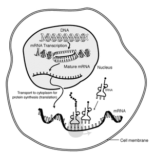
Protein synthesis has many steps. First, a copy of the DNA (deoxyribonucleic acid) is made in a process called transcription, then the copy is used to make a protein, in translation. Proteins are essential to life. The first step of translation is for a section of DNA, known as a gene, to be copied by an enzyme, in the nucleus. The copy is called messenger RNA, often abbreviated as mRNA. RNA has a few main differences form DNA: It is only single stranded, and the base uracil replaces thymine. 

After the copy is made, the mRNA leaves the nucleus and travels to the cytoplasm. Then, translation beings - the mRNA bonds with a ribosome, which will make a protein. The ribosome reads the first three bases called a codon, and determines which amino acid corresponds with that sequence. Each amino acid that is determined by the codon is read by the ribosome. Amino acids are bonded together and when the mRNA is done being translated, the amino acid chain fold up to become a protein. 

The gene expression and regulation lesson was particularly hard for me, but it was the one that I found the most interesting. The basic questions of that unit was: Why do genes appear in the correct places and at the correct times? Why don't we have eyes on our feet and toes on ours heads? And the answer was gene expression and regulation. 

Gene expression is the process of a gene being used to produce a gene product of the phenotype, basically what gene is expressed in the person. Gene regulation is a mechanism used by cells to increase or decrease the expression of a certain gene.

Another essential concept in this unit was Mutations. Although the connotation of the word mutation notes otherwise, mutations are generally very small and can have little to no effect. Is biology a mutation is a change in the DNA code. Mutations are happening almost constantly in our body. There are two main types of mutations - substitution and frameshift mutations. Substitution is when one nucleotide is substituted for another. There are two types of frameshift mutations - insertion and deletion. Insertion is when an extra base is put in and deletion is when a base is taken out.

The effect of the mutation is truly determined by where the mutation is placed. Suppose a harmful mutation to create a STOP amino acid, is placed at the front of the amino acid sequence - then the harms would be devastating. But, if the same mutation is placed near the end, then the amino acid sequence is still changed, but on a much smaller scale, thus it would not be that harmful.

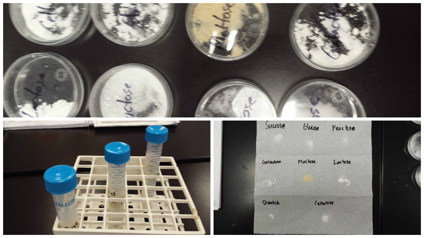
One of my strengths in this unit were the labs - They really helped em visualize and understand the processes. We did a DNA extraction lab as well as a protein synthesis lab, among other things. These labs allowed me to get a full understanding of the concepts. The process of doing the lab, whether the product came out good or not, helps me understand concepts much better than reading or watching a vodcast.

One of my weaknesses in this unit were the vodcasts. Some of the vodcasts were hard to fully understand, but I was able to ask questions to my group and do the labs to make up for it. The vodcasts were especially hard because diagrams were confusing. I was not able to follow some of the diagrams, but when we did them on the board in class, I got the concepts.

Overall my growth as a learner in this unit has become tremendous, mainly because of a VARK questionnaire that I took at the end of my last unit. It told me that I was a better visual learner, and I have always known that I always understanding best when actually doing something. I feel that I have really learned what helps me understand certain concepts and how I should study in the future for science, or for any other subject. This will help me throughout my years of learning, especially as finals week comes nearer.

This unit has also taught me about how to react well to setbacks. During the DNA extraction lab, at first, I was not able to extract DNA properly, due to a mistake in our procedure. But instead of getting mad at myself, I kept my head held high and redid the lab in the correct way. I was able to finish the lab before class ended and thus, I learned how to recover form a small setback in a lab.

This unit also really helped me learn how to collaborate with others in my group. During most of my previous units I understood most of the vodcasts and there was no need, really, to discuss properly with my group about it. But in this unit, since I did not understand the vodcasts completely, I needed to collobarate with my group to make sure I got the concepts down.

Image result for substitution mutations


Monday, December 12, 2016

Protein Synthesis Lab

In this lab, we asked the questions, "How does the body produce proteins and What kinds of mutations cause the greatest damage to the structure of a protein?" To answer these questions, we followed the steps of protein synthesis, throughout transcription and translation.

Protein production can be sorted into two main steps, with multiple substeps. First, a copy of the DNA (deoxyribonucleic acid) is made in a process called transcription, then the copy is used to make a protein, in translation. Proteins are essential to life. The first step of translation is for a section of DNA, known as a gene, to be copied by an enzyme, in the nucleus. The copy is called messenger RNA, often abbreviated as mRNA. RNA has a few main differences form DNA: It is only single stranded, and the base uracil replaces thymine. After the copy is made, the mRNA leaves the nucleus and travels to the cytoplasm. Then, translation beings - the mRNA bonds with a ribosome, which will make a protein. The ribosome reads the first three bases called a codon, and determines which amino acid corresponds with that sequence. Each amino acid that is determined by the codon is read by the ribosome. Amino acids are bonded together and when the mRNA is done being translated, the amino acid chain fold up to become a protein.


"Protein Biosynthesis." Wikipedia. Wikimedia Foundation, n.d. Web. 12 Dec. 2016. <https://en.wikipedia.org/wiki/Protein_biosynthesis>.

Of the mutations that we tested in this lab, I was able to conclude that each mutation could be nearly as harmful as every other mutation, depending on of the situation, the code, and when the mutations occur. We tested three types of mutations - substitution, insertion and deletion. The effect that substitution caused varied - only being extremely harmful when placed in a certain position. Otherwise, substitution is generally harmless. Insertion and deletion followed a similar pattern. When put near the start of the sequence, the change did quite a bit of damage, but when placed at the end, not much damage occurred.
substitution


"What Types of Mutation Are There?" Facts. The Public Engagement Team at the Wellcome Genome Campus, 25 Jan. 2016. Web. 12 Dec. 2016. <http://www.yourgenome.org/facts/what-types-of-mutation-are-there>.

When I chose my mutation I chose to replace the first G with the First T, as I wanted to see what happened if one of the early letters was changed. What ended up happening was that, the stop codon (UAA) was the second amino acid in the sequence, after MET, the start codon. This would have a devastating effect, especially if the protein was important, as it would not be able to make the protein. This shows why the place that the mutation is located can have a really big effect. Earlier in the lab we did a mutation at a different place in the same sequence and there was not a single effect on the amino acid sequence.
Image result for mutation

"What Types of Mutation Are There?" Facts. The Public Engagement Team at the Wellcome Genome Campus, 25 Jan. 2016. Web. 12 Dec. 2016. <http://www.yourgenome.org/facts/what-types-of-mutation-are-there>.

Because proteins are so essential to life as they play a part in almost every activity we do, mutations can sometimes be extremely harmful, especially if it greatly alters the amino acid sequence formed by the mRNA sequence. But it is also important to note that mutations happen often on a daily basis, and that most mutations are not as harmful. One example of a disease caused by mutations is phenylketonuria, abbreviate PKU. PKU is an incurable, chronic disease, that although rare, can cause great damage to the body. It is a birth defect that causes an amino acid called phenylalaline to build up in the body, which can lead to brain damage, intellectual disabilities, behavioral symptoms or seizures. PKU is an autosomal recessive disease caused by a mutated gene for the enzyme phenylalanine hydroxylase (PAH), which converts the amino acid phenylalanineto other essential compounds in the body.

Image result for phenylketonuria
"Phenylketonuria." U.S. National Library of Medicine. National Institutes of Health, n.d. Web. 12 Dec. 2016. <https://ghr.nlm.nih.gov/condition/phenylketonuria>.

Friday, December 2, 2016

DNA Extraction Lab

In this lab, we asked the question, "How can DNA be separated from cheek cells in order to study it?" We collected information about the three main steps of DNA extraction. Homogenization, lysis, and precipitation. We found that DNA from cheek cells could be extracted and studied if the correct steps are used in the correct order. The correct steps the proper order are as follows:
  1.  Measure 2.5mL of Gatorade into a paper cup
  2.  Scrape both sides of the inside of your checks using your teeth.
  3.  Vigorously swish in your mouth for 30 seconds.
  4.  Spit solution back into cup
  5.  Add a tiny punch of salt
  6.  Carefully poor solution into a test tube about 1/3 to 1/2 of the way up
  7.  Add 10 drops of detergent/soap.
  8.  Add 5-10 drops of your enzyme (pineapple juice)
  9.  Let sit for 5 minutes and record observations.
  10.  Tilt the tube at an angle and slowly add cold alcohol along the side of the test tube. You do not want the two layers to mix. The amount added should be about the same as the Gatorade mixture.
  11.  Collect the DNA and alcohol and carefully, with a transfer pipette, and place in a microcentrifuge tube. Do your best to only transfer DNA and Alcohol to your tube.
  12. Cover the tube with your thumb. Carefully invert 6 times. Be careful not to shake too much. You do not want soapy bubbles to form.
  13. Wait for another 5 minutes and record observations. 
Evidence from Experiment: During our first trial we switched steps 11 and 12, and thus we were not able to extract DNA, but when we did these steps in the correct order, we were able to extract DNA.

Reasoning: This evidence supports our claim, because during the first trial, which failed, the two layers, of alcohol and Gatorade, mixed and thus we were not able to separate DNA. However during the second trial which succeed we only inverted the tube after separating DNA, and thus was able to get the DNA.

Possible Error 1: The first error that we made was switching the order of the steps. Part of our lab was to put random steps in the correct order. We ended up switching steps 11 and 12, thus inverting the tube 6 times (and mixing the alcohol and Gatorade solutions), before separating the DNA. The effect of this error on the overall results was the fact that we were not able to get the DNA separate and the end of the experiment.

Possible Error 2: That was the only error we made in the experiment, but a hypothetical error could have been not adding the alcohol while the test tube was tilted. The effect of this would have been the same as the effect in PE 1, as the DNA would not separate at the end of the experiment.

This lab was done to help us understand the process of DNA Extraction, including the 3 main steps - homogenization, lysis and precipitation. I can relate this lab to the vodcast about "Your Genetic Code." The concepts from that vodcast, really helped me to do this lab, and my overall understanding of DNA was solidified through this lab process. The fact that we first messed up and then fixed our mistakes, really helped me understand some of the important concepts, like the enzymes breaking down the DNA.

From this lab, I learned the correct process of DNA extraction, and now I could extract DNA from any cell. This would be helpful if I wanted to study DNA, especially discrepancies in genetic variation, in the future as a scientist. Another outcome I learned from this lab is to slow down. At first, I though for sure that the first procedure we came up with was correct, but in hindsight, I feel like if I thought about it a bit more, I would have been able to see the mistake and correct it before we started the experiment. This teaches me to double check everything I do, and most importantly, slow down.


Sunday, November 27, 2016

Unit 4 Reflection

Coin Sex Lab


  • What did you do in this lab? How did coins serve as a model for genetics concepts?
  • Compare your expected results with actual results in your dihybrid cross simulation. To what can you attribute these results? (ie, make a claim and back it with reasoning).
  • What is the limit of using probability to predict our offspring’s traits?
  • How does understanding this relate to your life? Be specific

In this lab we predicted the outcomes of different types of crosses, by flipping coins to get phenotype. Each coin represented an gene, such as hair color, each side of the coin, heads and tails, represented alleles, such as blonde hair color or brown hair color.


We worked in partner groups and each person was given either 1 or two coins. Each person represented a parent and each person's coins represented the genes of each parent. When one person flipped their coins in the air, it represented meiosis.


The process of putting together the coin flip outcomes and showing them in terms of phenotype , that process simulated, sex or recombination.


We simulated multiple different crosses, including mono hybrid, and dihybrid crosses. For our dihybrid cross simulation, we received different than expected results. Because we only flipped these coins 10 times, the results were a bit skewed due to lack of testing, but the main reason was that the resulting phenotype of any cross at the end is, - random.

This creates a limit for using probability to predict our offspring's traits. Although we may be able to assume our offspring have a better chance of getting a specific trait, in the end it is all random, so we can never be 100 percent sure that our offspring will receive a certain trait. This is mainly because of Gregor Mendel's Law of Independent Assortment.

This lab relates to my life, because it educated me a lot about how traits get passed on and how we cannot predict it. All my life, I have always assumed that I have gotten my traits from just my mom and my dad, but now I know that is not true. Traits can skip generations, because of the randomness in Mendel's law of Independent Assortment. I also know now that I cannot predict what traits anybody's offspring might have.

In this unit we learned all about genetics, sexual vs asexual reproduction, cell growth and cell division, inheritance - autosomal vs X Linked, etc. We also learned about dominance vs recessive, and how sometimes there are exceptions and complications to every rule. In the instance of dominant vs recessive, some of the exceptions were codominance and incomplete dominance. We learned about mitosis, meiosis, and spent a lot of time comparing the two, their similarities and their differences.

One of my strengths in this unit was punnent squares. It was generally really easy for me to visualize the results, however I did not always get expected results in the punnent square or in any of our labs. That was probably one of my greatest weaknesses - assuming something would happen without testing it first. Time and time again in this unit my hypothesis was proven wrong when I actually tested out my work. Another one of my weaknesses was understanding the law of Independent Assortment. I originally though that the phenotype of the parents is automatically passed on to the offspring, and I had a hard time understanding that the recessive allele, the one parents are carriers for but don't exhibit, has the same chance to be passed on to the offspring as the dominant alleles.

Our genetics infographic really helped solidify my understanding of some concepts. The research portion, especially finding images allowed me to look at different sources and see different persepctives about concepts. I feel like this was one of the units I understood the best, because of the infographic.

I would love to learn more about genetic exceptions and complications, especially mutlifactorial disorders. Is there a way to mask the multifactorial disorder, and how badly can the environment affect a persons to change their behavior? Is there a way to predict multifactorial disorders, and what are ways to fix them?

My VARK Questionnaire scores were:

  • Visual: 18
  • Aural: 15
  • Read/Write: 16
  • Kinesthetic: 15
You have a multimodal learning preference. (VARK)

Most of the above results did not surprise me, as I have always been able to grasp ideas in many different ways, however, I was extremely surprised that the best way of learning for me was visual. I always thought physical activity or reading was my best way of grasping knowledge.

Because of this, I am going to use many videos, and diagrams to quiz myself in reparation for my upcoming test. I generally read textbook, articles online in preparation for tests, but hopefully my new idea of visual learning will help me better.

Image result
Dihyrbid Cross

Image result for law of independent assortment
The Law of Independent Assortment


Sunday, October 30, 2016

Is Sexual Reproduction Important?

In this blog post I will answer and provide supporting evidence for the question, Is Sex Important? And the short answer is yes. I will delve further into the reasoning and evidence for this answer in four main categories. The benefits of reproducing sexually, the costs of reproducing sexually, the benefits of reproducing asexually and the costs of reproducing sexually. Finally I will summarize all my points and justify why reproducing sexually is the better and safer way to reproduce.

Benefits of reproducing sexually:

Because of the concept of natural selection, genetic variation is crucial to survival. Without genetic variation, there is no evolution, and thus no survival. Genetic variation comes from two main sources mutation and sex.

Sexual Reproduction Benefit 1:

Sexual reproduction is the more sophisticated form of genetic variation. Mutations are random changes to information contained in genes, and is more primitive than sexual reproduction. Mutations only arise from errors made by the cells genetic copying machinery.

Make no mistake, sexual reproduction only produces combinations of genes that already exist, whereas mutation creates altogether new genes, and thus is necessary for generating the raw material of evolution. According to Dr. Tatiana, "Without mutation, evolution would grind to a halt"

Sexual Reproduction Benefit 2: 

Although mutations may be necessary, mutation alone is not enough. When organisms evolve to give up sex, and reproduce asexually instead. (I will explain more about the benefits and harms of asexual reproduction in the following sections, thus is just a comparison between sexual and asexual reproduction)

When an organism reproduces asexually, the differences between a parent are a child, are only due to mutation. According to Dr. Tatiana, "At first, these organisms often flourish. But their glory is fleeting. For reasons that remain mysterious, the loss of sex is almost always followed by a swift extinction."

Although there is one exception to this rule, the need for new combinations of genes that already exist are necessary, and without sexual reproduction, organisms cannot flourish. Most organisms need both sexual reproduction and mutations to properly survive and evolve.

Sexual Reproduction Benefit 3:

The final benefit of sexual reproduction is the ability to reproduce diverse offspring. I previously touched about this at the end of my first point, when I mentioned that in asexual organisms, mutations account for every difference in an organisms genetic makeup.

However, I never applied the importance of this to why sexually reproducing diverse offspring is good and why only mutational differences are bad. I will get to the cons of mutations differences in the "Asexual Reproduction costs," but for now, I will explain why having diverse offspring is helpful.
When having diverse offspring, organisms can ensure that the offspring will always be a combination of the two parents' genes and will never be the exact same, whereas in asexual reproduction, you can never control when mutations should happen.

For example if there is a poison in the environment, and for an organism to survive they need a particular gene. Asexual reproduction might take centuries for that organism to undergo a mutation that gives them that gene, and by the time that might happen, the organism will probably be extinct.
According to Dr. Tatiana, "More often than not, however, some individuals are fortunate and have a gene to resist the poison. Since these individuals are the only ones fo survive and reproduce, the genetic makeup of the population will shift to one where everybody is resistant.

Basically what this means is that sexual reproduction allows organisms to create offspring with genetic variation that might have the gene that resists the poison - If an organism does not have the necessary gene, but their mate does, then the offspring will have a good chance at getting the necessary gene and surviving the poison. Although this is not guaranteed, sexual reproduction allows organisms a better chance to survive the poison.

Sexual Reproduction Benefit 4:In my last point I discussed, how sexual reproduction would give organisms the ability to protect against a poison already spreading through the environment. They did this by mating with other organisms of their species that had a protective gene, to create an offspring that would survive.
This point is similar but it talks about how the genetic variations that sex provides with every reproductions, helps species and organisms survive - you could call it a contingency plan of sorts for the organism to almost always live on.

Genetic variations in every organism, allow each an every organism to have certain genes that protects them against almost any situation. According to Dr. Tatiana, "Monocultures are vulnerable to disease because all the individuals are the same clone," however, sexually reproducing organisms are not as vulnerable, because at least one of the organism should have the necessary genes to protect against anything.

This ensures that organisms that use sexual reproduction, will always survive any attack or poison, and live long, as all of them have different genetic makeups. The only reason that a sexually reproducing organism to go extinct is a drastic change in the habitability of their environment.

Sexual Reproduction Cost 1:

Sexual reproduction is hard to accomplish, and sometimes is a dangerous process. As opposed to asexual reproduction, sex requires two mates. This means that organisms have to seduce each other in order to reproduce sexually. 

According to Dr. Tatiana, "the competition for mates is often exceedingly stiff." Creatures might need to wear gaudy costumes, sing for hours on end, or perform prodigious feats to get a mate. Worse, the competition for mates is often at odds with survival. If you are a bird, flaunting an enormous tail may make you quite the cock among hens, but it also may make you lunch for a cat.

Sexual Reproduction Cost 2:

Another problem in the process of sexual reproduction is the time it takes, not only to find a mate, but to give birth to another organism. Although the time it takes to give birth to the organism may differ among different species, the time it takes to find a mate is never quick nor easy, as explained in my last point.

Asexually reproducing organisms do not need to find a mate, thus the time it takes for them the produce offspring is significantly shorter than the time is takes a sexually reproducing organism to do the same. 

Additionally the chance to make clones, or produce multiple offspring, although possible is very rare in sexually reproducing organisms. In asexually reproducing organisms, there is a much higher chance is producing multiple offspring each time. 

Furthermore, in sexually producing organisms, for example humans, there is usually a time limit between reproducing multiple times, whereas in asexually reproducing organisms, there is usually no time limit. Thus populations of asexual organisms usually grow at a much faster rate, than sexually reproducing organisms.

Asexual Reproduction Benefit 1:

Asexual organisms can reproduce faster and more effectively than sexually reproducing. According to Dr. Tatiana, "Sex may be fun, but cloning is much more efficient. All else being equal, an asexual female who appears in a population should have twice as many offspring as her sexual counterpart."

The reasoning for this is because in a sexual population, for example the human population, each female must have two children for the population to stay the same size. To cause the population to grow, each female in the population needs to average more than 2 children. If females average less than 2 children in a sexually reproducing population, the population will automatically shrink.

However, if an asexual female reproduces, it ensures that the population size will be maintained. If the organism reproduces more than once, then the population will automatically grow.

Asexual Reproduction Benefit 2:

Asexual reproduction is easy and does not take much time, while still allowing asexually reproducing species to produce multiple mates.

In asexual reproduction there is not necessity to find a mate, significantly lowering the time and difficulty it takes to reproduce. Additionally the reproduction itself does not take as much time as sexual reproduction. 

Another benefit that comes with the time factor is the quantity. Because of the shortened time and small amount of difficulty, asexually reproducing species can reproduce many more offspring in a small amount of time that sexually reproducing species can produce in a lifetime.

Asexual Reproduction EXAMPLE - Benefit 3:

One perfect example of an asexually reproducing species that has lived long for 85 million years is the philodina. It has not needed a single mate, and has produced many offspring.

Asexual Reproduction Cost 1:

Asexually reproducing organisms have no genetic variation. All the offspring of asexually reproducing organisms are genetically identical, aside from mutations that take place.  This means that every asexually reproducing organism is resistant to change.

This is one of the primary reasons that asexually reproducing species, according to Dr. Tatiana, "swiftly go extinct." If there is a poison, if one asexual organism in a species does not have a gene to fight the poison or virus, then none of the species will survive.

However, in sexually reproducing organisms, like humans, certain organisms have different genes that give them the ability to fight off viruses and diseases (as mentioned in the first benefit of sexual reproduction), even if one of the organisms die because of the virus.


These are the reasons that most sexually reproducing organisms thrive in our society, while asexual reproducing species usually go extinct swift because of pathogens that they are not resistant to.

Questions and What you want to learn more about:

I really want to learn more about the how pathogens affect sexually reproducing species versus asexually reproducing species.

I did not really understand why and how mutations happen, and what genes they specifically alter.


Tuesday, October 25, 2016

Unit 3 Reflection


In this unit, we learned all about cells - everything from their structure to their function(s) to their organelles. Some of the major sub topics covered in this unit were membranes, osmosis and diffusion, macromolecules found in the cell, the organelles in a cell, the history of cells, photosynthesis, and cellular respiration. In this unit, I really enjoyed doing all of the labs, especially the microscopic organism lab. The ability to magnify the organism, really solidified my learning, and was able to give me a deeper understanding.

Strengths and Weaknesses:

I really found myself to be good at understanding pictures under the microscope, and identifying key parts of the cells. However, I could never really get good pictures under the microscope, as my hands could never stay still. :(

Another strength I found in myself was my ability to interpret data. For the egg diffusion and egg macromolecule lab, this skill helped me understand why certain macromolecules were present in parts of the egg, and why egg grow and shrink in different types of liquids.

I think I am a better student than before this unit, especially because of the labs we did. The labs allowed me to test certain theories that I was not sure were true. They also provided me with a way to look closer and the cells. I also learned a life lesson from these experiences. There is always a reason. In this unit I refused to accept the answer, "That is just the way it is" and set out find why it is that reason. And I learned that there is always a reasonable explanation.

I want to learn more about the organelles of a cell and their specific functions. I want to look at the organelles closer in an electron microscope and see where the magic happens of photosynthesis. I always wonder about what we will find, when we keep looking closer at the cell.

Thursday, October 20, 2016

Microscope Organism Lab Analysis

The above diagram is of an amoeba cell. We were able to identify the nucleus, cell membrane, cytoplasm and pseudopods. One of the unique characteristics about this cell is that is has pseudopods to walk, and another is that they have different colors within the cell. One observation about the cell is that is has a cytoskeleton. The amoeba is a eukaryotic cell that is heterotrophic using their pseudopods to eat other cells.
The diagram above is of a euglena cell. We were able to identify chloroplasts, the nucleus, and the cytoplasm. However we could not identify the flagellum. One unique characteristic about the cell is that it has flagellum. Although it is very hard to see in the image above, flagellum are very rare in cells, but do exist in euglena. These are eukaryotic cells, but can be both autotrophic and heterotrophic.
The diagram above is of a bacteria cell. The coccus, bacilis, and spirilum are identified on the cell. The size of this cell compared to other cells, is extremely small, and an observation about the cell is that the bacteria are not in any particular formation - they are all just floating around. Bacteria are prokaryotic, heterotrophic cells. (400x)
The diagram above is of a spirogyra cell. On the cell the cell, the cell wall, cytoplasm and chloroplasts are identified. The cytoplasmic strands that hold the nucleus in place are one of the unique characteristics about the cell. One observation about the cell above, is that it has spiral chloroplasts, something that most plant cell's don't have. It is a eukaryotic, autotrphic cell. (400x)
The diagram above is an example of a plant ligustrum cell. It is an autotrophic eukaryotic cell. A main unique characteristic of the cell above is the epidermis cells surrounding it.  You can clearly see the chloroplasts, the blueish green places, where photosynthesis occurs. The veins of the cells are also very clearly visible. One observation about the image above is that all the cells are very compact and the cell walls are extremely thick, compared to other cells. (400x)
The diagram above consists of animal muscle cells, known as muscle fibers. We were able to label the nuclei (purple dots), the muscle fibers (long strands across cell), and straitons (the bands of fibers). One of the characteristics of the cell shown above that is unique is the fact that they are multinucleate cells. They have many nuclei fused together in one cell. An observation I made about the cell is that  the nucleus is not always surrounded in a certain way, it differs for each muscle cell. The animal muscle cell is a heterotrophic, eukaryotic cell. (Note: The microscope that took this picture was at x400 zoom, and after picture was taken, the image was zoomed in)


The diagram above is of cyanobacteria, also referred to as blue green algae. It is a bacteria, which means it is a prokaryotic cell and it is autrotrophic. One unique characteristic about cyanobacteria is that they were the first types of cells to perform photosynthesis. One unique characteristc seen in the slide is the formation of each cell of cynaobacteria. They live in clusters as opposed to a compact formation. One other observation is that the cells are never in a particular or similar shape, rather different types of round shapes. (400x)

Cell Characteristics

The autotrophic cells were usually green. They also all had chloroplasts as most of the autotrophic cells we observed were plants - and plants need chloroplasts to perform photosynthesis. Most of the autotrophic cells, although it may not have been visible, also had mitochondria, so they would perform cellular respiration.

The heterotrophic cells were mostly eukaryotic except for the bacteria, and most of them utilized some of their special characteristics to help them eat other cells - like the amoeba using its pseudopods.

All the eukaryotic cells had nuclei and all the prokaryotic cells lacked nuclei. Most of the prokaryotic cells were bacteria.


Tuesday, October 11, 2016

Egg Diffusion Lab Analysis

In this lab we tested the effect two solutions, one hypotonic, and one hypertonic would have on egg mass and circumference.

One of the eggs was placed in sugar water, while the other was placed in deionized water. The sugar water was a hypertonic solution, thus the egg shrunk in both mass and circumference. It had an average of -42.17% change in mass, and a -19.67% change in circumference, as it was going from high to low concentration - diffusion. The solute concentration was greater outside and the solvent concentration was greater inside. The cell tried to move into equilibrium, by using passive diffusion through the membrane, forcing solvent outside the cell, thus, causing the egg to shrink.

The other egg was placed in deionized water, a hypotonic solution, thus the egg grew. The movement of solvent inside the cell, caused the cell to grow by an average factor of 1.18% for mass and 1.94% for circumference.

Because the cell has to stay in equilibrium to maintain the balance of solute and solvent concentration, both the cell's internal and external environments change. This changes occur either through facilitated or passive diffusion through the cell membrane.

This lab demonstrates diffusion and hyoptonic and hypertonic solutions. It shows how a cell will always want to get into equilibrium despite the fact that it must change size to do so.

Fresh vegetables are sprinkled with water, so they can grow larger, while roads are sometimes slated to turn the roadside ice into water.

I would want to test what type of hypertonic and hyoptonic solutions cause the solute and solvent concentrations to change the quickest.


Friday, October 7, 2016


Egg Cell Macromolecule Lab Analysis

Clockwise starting from top left: Egg Membrane, Egg White, Egg Yolk, Pure Water

Egg Macromolecule Lab Analysis

In this lab we asked the question: Can macromolecules be identifies in an egg cell? And we found an overarching answer that macromolecules can be identified in all parts of the egg cell, whether it is the egg yolk, the egg membrane or the egg white.

Egg Membrane

Claim: The Egg Membrane tested positive for the macromolecule of lipid. We tested the presence of lipids in the membrane by mixing the egg membrane sample (shown in the top left of the picture above) with Sudan III, a solution that causes the sample to turn from red to orange if lipids are present.

The color change for the egg membrane in Sudan III
Evidence: When the Sudan III was mixed with the egg membrane, the sample did turn to a shade of dark orange. On a rating scale of 0 to 10, (0 = color of the negative control, 10 = very dark shade of orange), we ranked the color change to be 8.5 - The picture to the right shows the egg membrane mixed with Sudan III. This data clearly indicates that there is a strong presence of lipids in the egg membrane.

Reasoning: The bilayer of the cell membrane is made up of phospholipids - which are lipids what have a phosphate group in its molecule - nevertheless still lipids that are a major part of all membranes. Other types of lipids that are found in the egg membrane include cholesterol and glycolipids. Cholesterol maintains membrane structural integrity and fluidity, making it another essential part of the membrane. Glycolipids help maintain the stability of the membrane and act as a recognition site for certain chemicals to pass in and out of the cell membrane. All in all, lipids are a major and essential part to the function and structure of cell membrane, thus are present in the cell membrane. 

Egg White

Claim: The Egg White tested positive for the macromolecule of protein. We tested the presence of proteins in the egg white by mixing the egg white solution (shown in the top right of the picture at the very top) with sodium hydroxide copper sulfate, a solution which would turn any sample from blue to purple if the sample had proteins present. 
Proteins present in Egg White

Evidence: When sodium hydroxide copper sulfate was mixed with the egg white (shown in image to the right), the sample turned to a shade of dark purple. On the aforementioned rating scale, we rated the color change as a 6. The color change rating, compounded with the image shown to the right, clearly shows that there is a presence of proteins in the egg white.

Reasoning: The primary function of the egg white is to protect the yolk, also known as the nucleus of the egg cell. Proteins are found anywhere there is a membrane, because they (1) transport molecules and ions across the membrane as transport proteins, and (2) are attached to the lipid bilayer to help protect the cell as "integral membrane proteins." Some of the types of proteins present in the egg white are albumins, mucoproteins and globulins. The presence of proteins in egg whites does not just include transport and integral proteins, but is compounded by the fact that egg whites have 50 percent of the total protein in the egg. Thus, because the egg white's purpose is to protect the yolk, and proteins are found anywhere in the cell that serves as protection, proteins are present in the egg white solution.

Egg Yolk

Claim: The egg yolk tested positive for the macromolecule of lipids. We tested the presence of lipids in the egg yolk by mixing the egg yolk solution (shown in the bottom right in the very top image), with Sudan III, a solution which would turn any sample from red to orange if the sample had lipids present.

Evidence: When Sudan III was mixed with the egg white, the sample did turn to a shade of dark orange. On a rating scale of 0 to 10, (0 = color of the negative control, 10 = very dark shade of orange), we ranked the color change to be 4 - The picture to the right shows the egg yolk mixed with Sudan III. This data indicates that there is a presence of lipids in the egg membrane.

Reasoning: Egg yolk has both cholesterol and phospholipid contents. Cholesterol is a type of lipid that is present in the egg yolk, which helps protect the egg yolk, which is the nucleus of the egg cell. Phosvitins are also a type of protein present in the egg yolk, and they are important in getting calcium and iron to the nucleus. LIPIDS are found wherever there are membranes, as they are essential for any protection inside the cell. As the yolk is the nucleus, it contains a nuclear membrane, and thus lipids will be present in the nucleus.

Possible Errors

While our hypotheses were supported by our data, there may have been some possible errors during the steps of the experiment. One of the possible errors is the amount of solution placed into the egg yolk, egg membrane or egg white. If there was too much solution, whether it was Sudan III, sodium hydroxide copper sulfate, iodine, or benedicts, the effect of the solution may have been exaggerated. If there was too little solution, the true effect and potential of the solution may not have been shown. Although we tried to set a specific number of solution drops to limit the possible error percentage, no one can account for how much one drop is, how many drops may have accidentally be put in.

Another possible error could have been the mixing of the solution and the sample. One person may have mixed the solution and sample extremely well, clearly showing the color difference and properly telling us what macromolecules were present in each of the samples, however others may have recorded their data without properly mixing their solution and sample. If this did happen, the effect of the solutions on the samples would not have been shown, and would give us incorrect data to analyze. However the possibility of this is minimum. Although some people may have mixed the solution and sample more than others (ex. more time spinning the tubes), the procedure told us to mix it well, and the couple extra mixes would not provide too much of a data difference.

Two recommendations that I would give when doing this experiment in the future is to put the exact amount of solution (that should be poured) into a separate petri dish, that way groups can simply pix it up, and dump all the solution into the sample. This would prevent any errors of the quantity of drops from taking place. The second recommendation that I would give is to give the exact number of seconds a student should spend mixing, or the number of times a student should spend mixing, thus eliminating any possibility of excess or insufficient mixing.

Practical Applications

This lab was done to demonstrate the macromolecules that are present in all parts of the egg cell. From this lab I learned that every part of the egg cell, whether it is the egg yolk, egg membrane, or egg white, has at least one, if not multiple, macromolecules present. This helps the understand the concepts of macromolecules - especially the structure and function that each macromolecule serves in each part of the egg cell. This also helps me better understand the importance of macromolecules, as well as what each part of the cell does. With better knowledge of macromolecule function, I can quickly relate to and understand different functions of different cells. Based on my experience from this lab, I can eat much better. I understand that the egg yolk is mainly a source of cholesterol and saturated fats, both of which are not extremely good for your body, and that egg whites are the egg's main source of protein. This has allowed be to shift my morning meals from being full egg omelets, to solely egg white omelets, something that will hopefully help me put on more muscle, and still stay very healthy.

Thursday, September 22, 2016

Unit 2 Reflection

In this unit, we learned the basics of Miniature Biology, attempting to answer the essential unity question, "How does increasing molecular complexity serve as building blocks of life." We learned about atoms, elements, compounds, bonds, the Big 4 Macromolecules, and enzymes. Atoms are the basic building blocks of life and are made up of 3 particles, protons which are positively charged, neutrons which are neutral, and electrons, which are negatively charged. Protons and neutrons together form the nucleus, and electrons are circling the nucleus. We learned about ionic bonds, when an atom gains or looses an electron, covalent bonds, in which electrons are shared between atoms, and hydrogen bonds, that hold molecules together due to slight attractions of positive and negatively charged regions.

The Big 4 Macromolecules, were Carbohydrates, Lipids, Proteins and Nucleic Acids, molecules that ALL cells are made up of.  Each has a different characteristic, type, function, and are found in different places. For example, Carbohydrates are used as our main source of energy, and are broken down by mitochondria to make ATP. In a nutshell, enzymes are biological catalysts, which speed up chemical reactions, converting substrates into products. They can be affected by both pH and temperature. I really enjoyed and understood learning about bonds and macromolecules. But I really struggled when learning about enzymes and the basic function they serve - speeding up reactions.

Throughout this chapter, I really learned the importance of perseverance. At first glance, I did not understand enzymes at all. I read tons of information online and watched videos to understand the process and function of enzymes, and how specifically it speeds up the chemical reaction. Another skill I learned is too re-watch the vodcasts twice, once just to understand and to take notes the second time. This really helped me, as I could understand the vodcast on the first go and then solidify my knowledge when taking notes.

I really want to learn more about the four macromolecules and their effect on the human body. More specifically, I want to learn what each carbs, lipids, proteins and nucleic acids do to your body, especially when you are a teenager. Also, what type of carbs, and proteins should you get your energy from, and how this differs for different kids,


Friday, September 16, 2016

Sweetness Lab


Image result for saccharides
Three types of saccharides we used in this lab - Poly, Di, and Mono saccharides

In this lab we answered the question, "How does the structure of a carbohydrate affect its taste(sweetness)? After testing the sugars sucrose, glucose, fructose, galactose, maltose, lactose, starch and cellulose, we found that the monosaccharides (Glucose, Fructose, Galactose) were sweetest type of carbohydrate, while disaccharides came in second, and polysaccharides came in third and last place. This is supported by our observations as the average score for the monosaccharides on a scale of 0 to 200 was 155, while the average score for disaccharides was 70, and the average score for polysaccharides was 15. It is corroborated by our research, as the one molecule in monosaccharides make them sweeter than the 2 or more in poly and disaccharides. Monosaccharides are also used in sweet foods and drinks such as milk and sweet fruits.

Monosaccharides and Disaccharides are used mostly for immediate energy in cells and organisms due to their 1 or 2 molecule structure, while Polysaccharides are converted into energy for later use. The longer the carbohydrate is, the more energy it has. Plants use the ones with more energy for long term use, while they use the lesser energy carbohydrates for immediate energy.

No, all testers did not give each sample the same exact rating, for these three reasons.

  1. Everybody has their own opinion about certain types of tastes. Some people are generous raters while others are very strict.
  2. Everyone has different taste buds, and they taste things differently. Some have a preference for bitterness or sweetness which might cloud their rating judgment.
  3. People might take in a bit more of a sample than others cause them to like it more or less. For example, if you taste a lot of something bitter and you don't like it, you will give it a worse rating than someone who tasted very little of it. And vice versa for sweet samples.
According to the National Library of Medicine, the tongue uses its taste buds to sense the foods, and transports the information directly to the brain, which tells us how the food tastes. Our sense of taste used to be a matter of survival, which could tell us if plants were poisonous or edible, but now it is just a matter of what our taste buds enjoy eating. Everybody has different taste buds which might cause them to think that a food is more salty and savory, that sweet and sour. This is another reason to explain why all of the tasters ranked each sample differently, though they all got similar results.


Tuesday, September 6, 2016

Jean Lab Conclusion

Jean Lab

1st Lab for Scientific Method

Different Text Colors Used to Indicate Different %'s of Bleach in Data Results


In this Jean Lab performed in Mr. Orre's Classroom at Saratoga High School, we tested the effect of bleach on both the color and fabric damage of bleach, asking the question, "What concentration of bleach is best to fade the color out of new denim material in 10 minutes, without visible damage to fabric?". Using data recorded during the course of the experiment, we as a group, found out that the denim jeans squares with 50 percent concentration of bleach had the best cumulative result of more color removal and less fabric damage. Our evidence showed results as follows: The 3 denim squares soaked in 100% water (no bleach), had an average rating of 0 out of 10 for both color removal and fabric damage. On the rankings it turned out to have a cumulative score of 6 (5th place for color removal and 1st place for fabric damage), finishing tied for 3rd place. The 3 denim squares with a 12.5% concentration of bleach, got rated for an average of 3 out of 10 for color removal and an average of 2.33 out of 10 for fabric damage, with a cumulative score of 6 (3rd place for both color removal and fabric damage), finishing tied for 3rd place. The 3 denim squares with a 25% concentration of bleach had an average rating of 2.66 out of 10 for color removal and an average rating of 6 out of 10 for fabric damage, totaling a score of 9 (4th place for color removal and 5 place for fabric damage), finishing last at 5th place. The 3 denim squares with a 50% concentration of bleach, averaged a rating of 3.66 for color removal and 1 for fabric damage, with a cumulative score of 4 (2nd place for both color removal and fabric damage), finishing with a first place ranking. The 3 denim squares soaked in 100% concentration of bleach averaged a rating of 5.6 for color removal and 3 for fabric damage, totaling a score of 5 (1st place for color removal and 4th place for fabric damage), finishing at 2nd place. This evidence supports our claim that 50 percent concentration of bleach was the most effective to remove the most color with the least fabric damage, because it ended with a combined score of 4, finishing in 1st place against all the other concentrations, as it had the second least fabric damage and the second most color removal. The 1st place's in each category respectively, had extremely bad scores in the other category, as shown in the data table below.

The data table is listed below, with the final scores on the side. If you add the final scores for each concentration % respectfully, you will get the final rankings.

Concentration of Bleach (%)
Color Removal
Average
Score
0
0
0
0
0
5
12.5
3
3
3
3
3
25
2
2
4
2.66
4
50
4
4
3
3.66
2
100
8
6
3
5.66
1


Concentration of Bleach (%)
Fabric Damage
Average
Score
0
0
0
0
0
1
12.5
3
3
2
2.33
3
25
5
6
7
6
5
50
1
1
1
1
2
100
4
3
2
3
4

Our lab data contradicts the expected result of 12.5% percent being the most effective solution, mainly because of the pre-conditions to our denim jean materials. While setting up the lab, and following the procedure which we were given by our supervisor, Mr. Orre, we cut out squares from many different pairs of denim jeans, which all varies in color, as well as previous fabric damage. The experiment only exasperated both the color removal and the fabric damage of all the jeans. Because out procedure specifically told out us rate the color removal and fabric damage based on a general scale, rating the fabric damage solely on the appearance of the end product, instead of rating the effectiveness the bleach had on each square of denim, our results varied and contradicted the highly anticipated standard norm. While our hypothesis was greatly supported by our data, there could have been another possible error due to the size of the cut squares. Because of the tools we were provided with (scissors) it made the task of cutting perfect squares from ragged denim jeans extremely difficult. Our squares were not perfectly cut or sized, as most of them were ragged in the corners and varied in sizes. The sizes never varied more than a 1/2 inch, so it is not very likely that this created a huge effect or impact, but nevertheless, because of the varying size and shape, some of the denim squares may have absorbed more or less of the concentration in the 10 minutes, ergo creating another possible error that may justify the unexpected results we received. In conclusion, each of these two errors could have affected our results in drastic ways. The first possible error, because of the pre-given squares and the differences in color removal and fabric damage, caused our lab results to get thrown off, as some of the squares ended with more/less color removal and more/less fabric damage, entirely because they started our with different amounts of color and fabric damage. The second possible error, could also have accounted for our unexpected results, as different sizes and shapes could have absorbed different amounts of the bleach solutions, thus reducing the reliability and effectiveness of each bleach test. Due to these errors, in future experiments, I would recommend having a broader procedure, or letting the kid scientists choose procedures themselves, thus allowing them to account for any possible experimental errors in advance, before setting a procedure in stone. Another recommendation I would make for future experiments, is to have the exact same type of material for each group, or for each group to have their own materials set. For example, in this experiment multiple groups cut squares from many different jeans. In the future, we could assign one jean per table group, thus allowing tables to judge the effectiveness of the bleach based of the original color and fabric damage, rather than on a standard scale of 1 to 10.

This lab was primarily done to demonstrate understanding of basic science and biology practice,s including, but not limited to, the scientific method, conclusion writing practices, blog posts, following a procedure, accounting for errors, and most importantly, (in my opinion) working in conjunction with a group of other biologists, and sharing views while reflecting on the experiment. Although this lab was intended to solely give us an understanding of the way things will work in the biology classroom, and to get used to the lab setting and lab rules/parts, I felt like I improved in being in overall scientist as well. My group and I had to constantly be at the top of our game during class, as we were on a tight schedule to complete the experiment given our allotted class time. I felt we worked very well together, and succeed under pressure, ultimately completing the experiment and having an awesome time in the process. From this lab I learned the process of analyzing data and accounting for errors and applications, which really helped me relate to the concept of the conclusion writing format. In addition, I was also able to solidify my learning of the scientific method, and understand how to follow a given procedure to the fullest extent possible. Based on my experiences in the lab, I could now easily device and run a good science experiment, gathering, analyzing and concluding data. Anther situation that we could apply our learning in this lab to a research study. We now know the basics of writing summaries and conclusions for scientific experiments with lots of data, which gives us the basic building blocks and foundation to analyze bigger experiments such as research studies or surveys.


Copyright © 2014 Siva's Biology Blog. Designed by OddThemes | Distributed By Gooyaabi Templates